Common Genetic Diseases in Dog Breeds
Common Genetic Diseases in Dogs
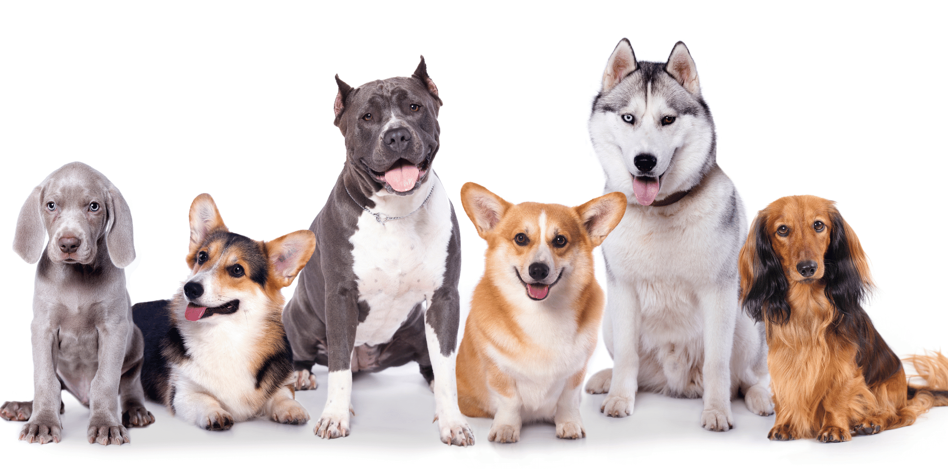
The genetics of dogs is a complex and fascinating field.
While selective breeding has led to the diverse range of breeds we see today, it has also predisposed certain breeds to specific genetic diseases.
This article explores some of the most common genetic diseases found in various dog breeds, emphasising the importance of awareness and responsible breeding.
Hip Dysplasia
Hip dysplasia is a condition that primarily affects the hip joint, which is a ball-and-socket joint.
In a healthy joint, the ball at the top of the thigh bone (femur) fits snugly into the socket in the pelvis, allowing for smooth movement.
However, in dogs with hip dysplasia, this joint doesn't develop properly, resulting in a loose fit and abnormal movement within the joint.
Over time, this improper fit can lead to various degrees of arthritis, discomfort, and decreased mobility.
Causes and Risk Factors
The primary cause of hip dysplasia is genetic.
- Diet: Overfeeding puppies, especially those of large breeds, can accelerate growth and potentially exacerbate the development of hip dysplasia. A balanced diet that promotes gradual growth is recommended.
- Exercise: Both excessive and insufficient exercise can be harmful. High-impact activities, particularly on hard surfaces or involving lots of jumping and running, can worsen the condition, while moderate exercise can help maintain healthy joint function.
- Body Weight: Overweight dogs have an increased risk of developing hip dysplasia due to the extra stress placed on their joints.
Symptoms
Symptoms of hip dysplasia can vary widely from one dog to another and may include:
- Limping or lameness in the hind legs
- Difficulty rising, jumping, or climbing stairs
- Audible sounds from the hip area during movement
- Decreased activity levels or reluctance to play
- Changes in gait, such as a "bunny-hopping" movement
Diagnosis
Treatment and Management
- Weight Management: Keeping the dog at a healthy weight can significantly reduce stress on the hips.
- Exercise: Low-impact activities like swimming can help maintain muscle strength without putting too much strain on the joints.
- Medications: Nonsteroidal anti-inflammatory drugs (NSAIDs) can be used to manage pain and inflammation. Other supplements, such as glucosamine and chondroitin, may also be beneficial.
- Surgery: In severe cases, surgical options such as total hip replacement or femoral head osteotomy may be recommended to improve joint function and reduce pain.
Prevention
Brachycephalic Syndrome
Brachycephalic breeds are popular for their distinctive appearance, characterised by broad, short skulls, flat faces, and short noses. This facial structure, while appealing to many, predisposes these breeds to a range of health issues collectively referred to as brachycephalic syndrome.
This condition is a direct result of the physical traits selectively bred into these dogs over generations. Bulldogs, Pugs, Boston Terriers, French Bulldogs, and Shih Tzus are among the breeds most commonly affected.
Key Components of Brachycephalic Syndrome
- Stenotic Nares: This condition involves the narrowing of the nostrils, which restricts airflow and makes it difficult for the dog to breathe through the nose.
- Elongated Soft Palate: The soft palate is the soft part of the roof of the mouth. If it is too long, it can extend into the airway and block airflow, leading to difficulty breathing, especially during exertion or excitement.
- Hypoplastic Trachea: Some brachycephalic dogs have a trachea that is narrower than normal, further restricting air flow to the lungs.
- Everted Laryngeal Saccules: With increased effort to breathe, the tissue within the larynx can become everted, obstructing airflow and exacerbating breathing difficulties.
Secondary Health Issues
The unique structure of brachycephalic breeds also leads to other health complications:
- Dental Issues: The shortened jaw structure of these dogs means their teeth are often crowded or misaligned, leading to dental problems such as increased risk of periodontal disease, tooth decay, and difficulty eating.
- Overheating: The compromised airway makes it hard for these dogs to pant effectively, which is a dog's primary means of cooling down. This inefficiency in thermoregulation makes brachycephalic breeds more susceptible to heat stress and heatstroke, especially in warm climates or during vigorous exercise.
- Skin Issues: The folds of skin on the face and body, characteristic of many brachycephalic breeds, can trap moisture and debris, leading to skin infections if not regularly cleaned.
Management and Care
- Avoiding Overheating: Limiting exercise during hot weather, providing plenty of shade and water, and using air conditioning can help prevent overheating.
- Weight Management: Keeping the dog at a healthy weight reduces respiratory and cardiac stress.
- Regular Veterinary Care: Routine check-ups can help identify and manage symptoms of brachycephalic syndrome early. Surgical interventions, such as widening the nostrils or shortening the soft palate, may be recommended in severe cases to improve the dog's quality of life.
- Special Diets and Feeding Methods: Feeding a diet suitable for the breed and using specially designed feeding bowls can help mitigate the risk of aspiration and ensure proper nutrition.
Ethical Breeding Practices
Progressive Retinal Atrophy (PRA)
Progressive Retinal Atrophy (PRA) is an umbrella term for a group of genetic disorders that affect the retina, leading to a progressive loss of vision and ultimately blindness in dogs.
This condition is inherited and is prevalent in several breeds, including but not limited to Cocker Spaniels, Labrador Retrievers, Poodles, and many others. PRA is particularly insidious because affected dogs may not show signs of vision loss until the disease is well advanced.
Understanding PRA
Types of PRA
The most common forms include:
- Rod-cone dysplasia (RCD), where the rods deteriorate first, followed by the cones.
- Cone-rod dystrophy (CRD), which starts with the loss of cone photoreceptor function, leading to day blindness and loss of detailed vision, followed by rod deterioration and night blindness.
Symptoms
- Increased difficulty seeing in low light or darkness, leading to reluctance to go into dark spaces or nighttime anxiety.
- Dilated pupils that reflect more light, giving their eyes a "shiny" appearance due to the increased effort to capture light.
- Eventually, visible clumsiness, bumping into objects, or hesitation in unfamiliar environments as the vision loss becomes more pronounced.
Diagnosis
Management and Treatment
Management strategies include:
- Maintaining a consistent environment to prevent injuries, as moving furniture can disorient a visually impaired dog.
- Using verbal cues to help guide and reassure the dog.
- Ensuring the dog is kept on a leash or in a secure area to prevent accidents when outside.
Research and Future Directions
The Importance of Responsible Breeding
The condition is commonly known as Von Willebrand's Disease (vWD), which is indeed analogous to hemophilia in humans.
It is the most prevalent inherited blood clotting disorder in dogs, affecting various breeds with notable frequency in Doberman Pinschers, Scottish Terriers, and Shetland Sheepdogs. This genetic disorder is characterised by a deficiency or malfunction of von Willebrand factor (vWF), a crucial protein in the blood clotting process.
Understanding Von Willebrand's Disease
Von Willebrand factor helps platelets to adhere to each other and to the walls of blood vessels, which is an essential step in the formation of a blood clot. When vWF is deficient or defective, the clotting process is significantly impaired, leading to prolonged bleeding times even from minor cuts or injuries.
The severity of the condition can vary widely among affected dogs, ranging from mild to severe.
Types of vWD
Von Willebrand's Disease is classified into three types based on the level of severity and the specific genetic mutations involved:
- Type 1 vWD is the most common and generally the mildest form. Dogs with Type 1 vWD have lower than normal levels of vWF.
- Type 2 vWD is less common and involves a qualitative defect in the vWF protein. Dogs with Type 2 have normal or reduced levels of vWF, but the protein is dysfunctional.
- Type 3 vWD is the rarest and most severe form. Dogs with this type have very little to no detectable vWF in their blood.
Symptoms
The symptoms of vWD can vary but often include:
- Excessive bleeding from the nose, mouth, or genital area
- Bleeding gums
- Blood in the urine or faeces
- Excessive bleeding following surgery or injury
- Prolonged bleeding during estrus or after giving birth in females
Many dogs with mild forms of vWD may never show symptoms unless they undergo surgery or sustain significant injuries.
Diagnosis
Diagnosis of vWD involves a combination of clinical signs and diagnostic tests. The most definitive test is measuring the level of vWF in the blood. Genetic testing is also available for some breeds, which can identify carriers of the disease or those likely to develop it.
Management and Treatment
While there is no cure for vWD, the condition can be managed with careful monitoring and preparation:
- Avoiding Certain Medications: Some medications, such as aspirin, can exacerbate bleeding tendencies and should be avoided in dogs with vWD.
- Surgical Precautions: For procedures that cannot be avoided, such as spaying or neutering, veterinarians may recommend infusion of blood products that contain vWF prior to surgery to reduce the risk of excessive bleeding.
- Emergency Preparedness: Owners of dogs with vWD should be aware of the signs of excessive bleeding and have a plan in place for emergency veterinary care.
Breeding Considerations
Responsible breeding practices are crucial for reducing the prevalence of vWD. Genetic testing allows breeders to identify carriers of the disease and make informed decisions to avoid producing affected offspring. By selecting against the genes responsible for vWD, breeders can gradually reduce the incidence of the disorder in future generations.
Von Willebrand's Disease is a significant genetic disorder affecting blood clotting in dogs, with some breeds being more predisposed than others. Understanding the condition, coupled with vigilant management and responsible breeding practices, can help mitigate the risks and improve the quality of life for affected dogs.
Degenerative Myelopathy
Degenerative myelopathy (DM) is a progressive, incurable neurological disorder that affects the spinal cord, primarily in older dogs. It leads to a gradual loss of coordination and strength in the hind limbs, eventually resulting in paralysis. German Shepherds, Boxers, Pembroke Welsh Corgis, and several other breeds are known to have a higher predisposition to this condition, suggesting a genetic component to its incidence.
The disease usually manifests in dogs between 8 to 14 years of age, but it can occur earlier or later.
Pathophysiology
Degenerative myelopathy initially affects the white matter of the spinal cord, particularly the part that transmits signals to and from the hind limbs. The disease causes the myelin sheath, which insulates nerve fibres and facilitates the rapid transmission of electrical signals, to deteriorate. This degeneration disrupts communication between the brain and limbs, leading to the clinical symptoms observed in affected dogs.
Symptoms and Progression
The progression of degenerative myelopathy occurs in stages:
- Early Stage: The initial signs may be subtle, including loss of coordination (ataxia) in the hind legs, difficulty standing up, and dragging of the feet. These may be mistaken for normal signs of aging.
- Intermediate Stage: As the disease progresses, symptoms become more pronounced. Dogs may exhibit significant weakness in the hind limbs, partial paralysis, and difficulty controlling bowel and bladder functions.
- Late Stage: The final stages of DM involve complete paralysis of the hind limbs. The disease may progress to affect the front limbs, leading to quadriplegia. At this point, quality of life considerations become paramount.
Diagnosis
Diagnosing degenerative myelopathy involves ruling out other conditions that can cause similar symptoms, such as spinal cord tumors, intervertebral disc disease, or chronic degenerative radiculomyelopathy (CDRM). A definitive diagnosis can be challenging but may be supported by advanced imaging techniques (MRI, CT scans), cerebrospinal fluid analysis, and, importantly, genetic testing. A specific mutation in the SOD1 gene has been identified in many dogs with DM, providing a genetic basis for the disorder.
Management and Treatment
While there is no cure for degenerative myelopathy, management focuses on maintaining quality of life:
- Physical Rehabilitation: Physical therapy, including exercises, swimming, and the use of mobility aids (wheelchairs), can help maintain muscle strength and mobility for as long as possible.
- Nutritional Support: A balanced diet, potentially supplemented with antioxidants, may support overall health, though no specific nutritional regimen has been proven to alter the course of DM.
- Comfort Measures: As mobility decreases, ensuring the dog has a comfortable, accessible environment and managing any secondary complications (like urinary tract infections due to incontinence) is important.
Research and Hope for the Future
Ongoing research into degenerative myelopathy includes studies on the genetic aspects of the disease, with the hope of developing gene therapy or other treatments that could modify the course of the disease. Understanding the genetic underpinnings has already led to the development of a genetic test, allowing breeders to identify carriers of the SOD1 mutation and make informed breeding decisions to reduce the incidence of DM in future generations.
Ethical Breeding Practices
With the availability of genetic testing, breeders have a powerful tool to minimize the risk of degenerative myelopathy in susceptible breeds. By testing breeding stock and considering the genetic status of potential sires and dams, breeders can work towards eliminating this devastating disease from the gene pool over time.
In conclusion, degenerative myelopathy is a challenging condition that requires a comprehensive approach to management focused on maintaining the affected dog's quality of life. Advances in genetic research provide hope for future interventions and highlight the importance of responsible breeding practices in reducing the prevalence of this disease.
Dilated Cardiomyopathy (DCM)
Dilated Cardiomyopathy (DCM) is a serious condition that affects the heart's ability to pump blood effectively, due to an enlargement and weakening of the heart's ventricles. It is particularly prevalent in certain dog breeds, including Dobermans, Great Danes, and Irish Wolfhounds, pointing to a strong genetic predisposition. The disease often progresses silently, meaning that by the time symptoms become noticeable, the condition is typically advanced.
Understanding DCM
Symptoms
As the condition progresses, symptoms may include:
- Weakness and lethargy
- Reduced appetite
- Weight loss
- Coughing
- Difficulty breathing
- Abdominal distension due to fluid accumulation
- Collapse or fainting
Diagnosis
Genetic Factors
Treatment and Management
- Diuretics, to remove excess fluid from the body
- ACE inhibitors, to reduce the heart's workload
- Beta-blockers or calcium channel blockers, to manage heart rate and blood pressure
- Positive inotropes, to improve heart muscle contractions
Prognosis
Breeding Practices
Canine Multifocal Retinopathy
Canine Multifocal Retinopathy (CMR) is an inherited eye condition seen in several dog breeds, including Mastiffs, Bulldogs, Boxers, and others. It is characterised by the development of multiple, raised lesions on the retina, which is the light-sensitive layer of tissue at the back of the eye responsible for capturing images and sending them to the brain.
Despite its potentially alarming presentation, CMR typically does not lead to complete blindness but can cause varying degrees of visual impairment.
Understanding Canine Multifocal Retinopathy
CMR lesions are caused by the abnormal development of the retinal pigment epithelium (RPE), the layer of cells beneath the retina that supports its health and functionality. These lesions can appear as gray, white, or orange areas on the retina and may vary in size. The condition often manifests in dogs between a few months to a few years of age, with the lesions becoming stable or even showing regression over time.
Symptoms and Detection
The primary sign of CMR is the presence of the distinctive lesions on the retina, which a veterinarian can observe through a detailed eye examination. Symptoms from the dog's perspective might be minimal or subtle, given the non-progressive nature of the condition in many cases. Owners might notice slight changes in their dog's vision, particularly in low light conditions, or a cautiousness in unfamiliar environments.
Diagnosis
Diagnosis of CMR is primarily through a comprehensive eye examination by a veterinary ophthalmologist. This may include direct visualisation of the retina using specialized equipment. Genetic testing is also available for some breeds, which can confirm the diagnosis if the responsible genetic mutation is identified.
Treatment and Management
There is no specific treatment required for CMR, as the condition is typically non-progressive and does not lead to complete blindness. Management focuses on monitoring the condition to ensure that it does not impact the dog's quality of life significantly. Regular veterinary eye exams are recommended to keep track of the lesions and assess any changes in the dog's vision.
Genetic Considerations
CMR is an inherited condition, with the mode of inheritance varying by breed. In some breeds, it is autosomal recessive, meaning that a dog must inherit two copies of the mutated gene (one from each parent) to show symptoms of the disease. Genetic testing can identify carriers of the gene as well as affected dogs, providing valuable information for breeders to make informed decisions and reduce the incidence of CMR in future generations.
Implications for Breeders and Owners
For breeders, responsible breeding practices are essential to minimise the spread of CMR. This includes genetic testing of breeding animals and considering the results in breeding decisions. For owners, understanding that CMR typically does not lead to severe vision loss or blindness is reassuring. However, awareness and regular eye examinations are crucial to ensure any potential impact on vision is identified and managed effectively.
Canine Multifocal Retinopathy is a genetic condition affecting the retina, leading to lesions that may cause mild to moderate visual impairment. While it does not usually result in blindness, understanding and monitoring the condition are vital for the well-being of affected dogs.
Through responsible breeding and awareness, the impact of CMR can be minimized, ensuring affected dogs lead full and active lives.
Collie Eye Anomaly
The inherited eye condition commonly seen in Collies and related breeds, such as the Shetland Sheepdog, Australian Shepherd, and Border Collie, is known as Collie Eye Anomaly (CEA). CEA is a congenital condition, meaning dogs are born with it.
The severity of the condition can vary greatly among affected dogs, ranging from mild, which might not significantly affect vision, to severe, which can lead to blindness.
Understanding Collie Eye Anomaly
CEA affects the development of the eye, specifically the choroid, which is a layer of tissue supplying nutrients and oxygen to the retina, the light-sensitive layer at the back of the eye. The anomalies associated with CEA can include choroidal hypoplasia (underdevelopment or thinning of the choroid), colobomas (defects in the optic disc), retinal detachment, and atrophy of the optic nerve. These issues can compromise the eye's ability to process visual information, leading to varying degrees of visual impairment.
Symptoms and Detection
Many dogs with CEA do not show overt signs of visual impairment, especially in cases of mild to moderate severity. The condition is typically bilateral, affecting both eyes, though the degree of impact can vary between the eyes. Severe cases, particularly those involving retinal detachment or significant colobomas, can lead to noticeable vision loss or blindness.
CEA is usually diagnosed through an eye examination by a veterinary ophthalmologist. This exam can identify characteristic changes associated with the condition, even in very young puppies, which is crucial for early detection.
Genetic Basis and Breeding Considerations
CEA is inherited in an autosomal recessive manner, which means that a dog must inherit two copies of the defective gene (one from each parent) to be affected. Dogs with one copy of the gene (carriers) do not show symptoms but can pass the gene to their offspring. This genetic understanding allows breeders to screen their dogs for CEA through genetic testing, which can identify affected dogs, carriers, and those free of the gene.
Responsible breeders use these genetic tests to make informed decisions about breeding pairs to reduce the incidence of CEA in future generations. By avoiding the breeding of two carriers or an affected dog and a carrier, breeders can significantly decrease the prevalence of the condition in the breed population.
Management of Affected Dogs
There is no cure for CEA, but most dogs with the condition live relatively normal lives, especially if their case is mild or moderate. Dogs with severe CEA or those who become blind may require special care, including safe environments to prevent injuries and training to adapt to their limited vision.
Owners of dogs with CEA should have their pets' vision monitored regularly by a veterinarian to identify any changes or complications early. Additionally, understanding the condition's genetic nature can help owners make informed decisions about spaying or neutering affected dogs to prevent the transmission of the gene to future generations.
In summary, Collie Eye Anomaly is a significant inherited condition in Collies and related breeds, with a wide range of severity. While it can lead to blindness in severe cases, many affected dogs lead full lives with minimal impact on their vision. Through responsible breeding practices, including genetic testing and selective breeding strategies, the prevalence of CEA can be reduced, contributing to the overall health and well-being of these beloved breeds.
The Importance of Genetic Testing
The field of veterinary science has seen remarkable advancements in the realm of genetic testing, making it a powerful tool in the fight against inherited diseases in dogs.
This progress has significantly impacted how breeders approach the health and genetic integrity of their breeding programs. Genetic tests can now identify carriers of specific diseases, affected individuals, and those completely free of certain genetic conditions.
This capability enables breeders to make informed decisions, reducing the prevalence of genetic disorders in future generations of dogs.
The Role of Genetic Testing in Breeding Programs
Genetic testing offers several benefits in the context of responsible breeding:
- Identification of Carriers: Testing allows breeders to identify dogs that carry one copy of a gene for a recessive disorder. While carriers are typically healthy themselves, they can pass the disease gene to their offspring. Knowing a dog's carrier status helps breeders avoid mating two carriers, which would risk producing affected puppies.
- Prevention of Disease: By identifying and selectively breeding dogs that are free from specific genetic mutations, breeders can gradually reduce the occurrence of these diseases in the breed population.
- Informed Decision Making: Genetic testing provides breeders with concrete data about their dogs' genetic health, enabling them to make decisions based not on guesswork but on solid evidence.
- Preservation of Genetic Diversity: Thoughtful use of genetic testing can help maintain or even increase genetic diversity within a breed. By identifying carriers of rare genes, breeders can include these dogs in their breeding programs in a way that avoids producing affected offspring while preserving valuable genetic diversity.
How Genetic Testing Works
Genetic testing for dogs typically involves collecting a DNA sample from the dog, usually via a cheek swab or a blood sample. This sample is then sent to a laboratory specialising in veterinary genetic testing, where it is analysed for specific genetic markers associated with inherited diseases known to affect the breed.
Common Diseases Screened by Genetic Tests
The range of diseases that can be screened via genetic testing is continually expanding.
Some of the more common inherited diseases that breeders commonly test for include:
- Hip Dysplasia: Though largely influenced by environmental factors, genetic predispositions play a crucial role.
- Degenerative Myelopathy (DM): A progressive disease of the spinal cord, leading to paralysis.
- Von Willebrand's Disease (vWD): A blood clotting disorder.
- Progressive Retinal Atrophy (PRA): A group of diseases leading to blindness.
- Collie Eye Anomaly (CEA): An eye disorder that can lead to vision problems or blindness.
- Dilated Cardiomyopathy (DCM): A serious heart condition.
Challenges and Considerations
While genetic testing offers many benefits, it also presents challenges. One risk is the potential for overemphasis on genetic purity, which could lead to reduced genetic diversity if dogs are excluded from breeding programs based solely on their carrier status for certain diseases.
Breeders must balance the desire to reduce the incidence of genetic disorders with the need to maintain a broad genetic base within the breed.
A negative test result does not guarantee a puppy will never develop any health issues, as not all diseases are genetic, and not all genetic diseases can be tested for currently. Thus, while genetic testing is a powerful tool, it should be one component of a comprehensive approach to breeding healthy dogs.
The accessibility of genetic testing in veterinary science represents a significant advancement in the effort to breed healthier dogs. Responsible use of these tests allows breeders to reduce the likelihood of inherited diseases in puppies, contributing to the overall health and longevity of future generations.
Implications for Dog Owners
Owners of breeds predisposed to these genetic conditions should be aware of the signs and symptoms. Regular veterinary check-ups and a proactive approach to health can help in managing these diseases.
While many dog breeds are predisposed to certain genetic diseases, awareness and responsible breeding can help mitigate these risks. For dog owners, understanding these genetic predispositions is crucial in providing the best care for their furry companions. Advances in genetic research continue to provide hope for better management and treatment of these conditions in the future.
More Skin Tips.
CoreBodi